
Dr. Russell Blaylock: Aluminium, childhood vaccines and the rising rates of autism (Part 2)
In June, Dr. Russell Blaylock published a paper which describes aluminium’s neurotoxic properties and the connection between childhood vaccines which contain aluminium and autism spectrum disorder (“ASD”). “In this paper, I offer a well-dem
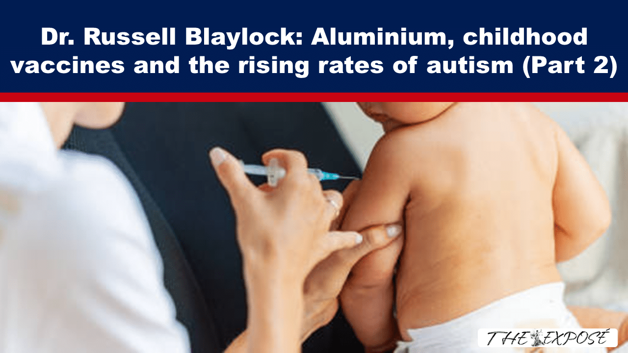
In June, Dr. Russell Blaylock published a paper which describes aluminium’s neurotoxic properties and the connection between childhood vaccines which contain aluminium and autism spectrum disorder (“ASD”).
“In this paper, I offer a well-demonstrated mechanism that would explain why a subset of children develop autism after vaccines,” he wrote.
Let’s not lose touch…Your Government and Big Tech are actively trying to censor the information reported by The Exposé to serve their own needs. Subscribe to our emails now to make sure you receive the latest uncensored news in your inbox…
Stay Updated!
Stay connected with News updates by Email
In June, Dr. Russell Blaylock published a paper on the connection between autism spectrum disorders and vaccines in the journal Science, Public Health Policy and the Law. We are republishing this paper in a series of articles. Although it is not overly technical, it does include some terms and concepts we may not be familiar with. By publishing it piecemeal, we are hoping our readers do not become overwhelmed by jargon that might be the case had they been faced with the entire paper at once. Also, it might give an opportunity for pause, to look up and familiarise themselves with terms as required.
You can read Part 1 HERE, where Dr. Blaylock provides an overview of the factors that contribute to a person developing an autism spectrum disorder. If you would like to read the paper in one sitting, you can do so HERE. Please note, we have not included the references noted in the paper as originally published. And we have made some minor edits to convert American English to British English and preferred stylisation, e.g. removal of Oxford commas.
Autism Spectrum Disorders: Is Immunoexcitotoxicity the Link to the Vaccine Adjuvants? The Evidence
By Russell L. Blaylock, as published by Science, Public Health Policy and the Law on 1 June 2025
“I coined the term ‘immunoexcitotoxicity,’ which describes the interplay between immune activation and excitotoxic neuronal injury.”—Russell L. Blaylock, Autism Spectrum Disorders: Is Immunoexcitotoxicity the Link to the Vaccine Adjuvants? The Evidence
[Note from The Exposé: Immune activation is the process by which the immune system initiates a response to eliminate foreign pathogens or abnormal cells. Excitotoxicity is a pathological process in which nerve cells (neurons) are damaged or killed due to excessive stimulation by neurotransmitters, primarily glutamate, the main excitatory neurotransmitter in the central nervous system (“CNS”). Immunoexcitotoxicity is a combination of immune activation and excitotoxicity.]
Excitotoxicity and Neurodevelopment
Excitotoxicity (Immunoexcitotoxicity)
Dr. John Olney discovered excitotoxicity in 1969. I knew Dr. Olney and visited his lab in the 1980s. Since his discovery, a whole array of new receptors and the physiology and pathophysiology of these glutamate receptors have been discovered. I suggested a link to the ASD and Attention-Deficit/Hyperactivity Disorder (“ADHD”) in a book I wrote in 1990. Initially, I suspected excitotoxicity contributed to autism spectrum disorders. My research into chronic traumatic encephalopathy (“CTE”) revealed a critical link between immune activation and excitotoxicity, leading me to identify immunoexcitotoxicity as a central mechanism in ASD. I called this link between the two systems immunoexcitotoxicity. Although I coined the term, I did not make the initial link. Furthermore, I discovered a link between the adjuvants commonly used in vaccines, such as aluminium, and excitotoxicity.
How Immune Activation Triggers Excitotoxicity
Immunoexcitotoxicity Answers Many Questions Not Answered by Other Mechanisms: Immunoexcitotoxicity During Neurodevelopment
Stimulating the immune system peripherally, especially repeatedly, will trigger brain excitotoxicity by a process of immunoexcitotoxicity. To understand excitotoxicity, one must understand glutamate receptor physiology, which is quite complex. In the newborn or small child, one must understand the effect of both pro-inflammatory cytokines and excitotoxins on neurodevelopment through their reaction with microglia. While microglia and astrocytes normally provide support to the neurons during brain development, in the face of inflammation, these cells are switched to a destructive mode. Many physicians, including paediatricians and obstetricians, lack this understanding.
Stimulating the systemic immune system (as with the flu, otitis media, or a series of vaccinations) will activate the microglia and astrocytes in the CNS, especially within the brain. This connection is made via pro-inflammatory cytokines traversing the blood-brain barrier, cytokine passage through the circumventricular organs (which contain only a partial barrier), and the cranial nerves connecting directly to the CNS (vagus and trigeminal nerves). During early birth, the blood-brain barrier (“BBB”) is immature and can allow the passage of toxic molecules and inflammatory cytokines, chemokines. The activation of the CNS glia is rather rapid (minutes) and can explain the shrill encephalopathic cry and sudden seizures sometimes seen in some children after vaccination, especially babies. It is not the pain of the injection, but an immune excitotoxic reaction affecting the brain. The covid injection will be worse in many ways, as the spike protein is deposited throughout the vascular system (endothelium), other organs and the CNS. It will act as an intense, continuous source of immune activation in microglia and astrocytes, resulting in immunoexcitotoxicity.
Impact on Neurodevelopment
Microglia, often referred to as the brain’s resident immune cells, play a nurturing and supportive role during normal brain development by maintaining homeostasis and promoting neural growth. Within the CNS, microglia are the main resident immune cells. Yet, macrophages can enter the brain and act like resident microglia. Except for special staining, these cells cannot be distinguished from resident microglia. Microglia can also migrate within the brain to sites of activation. While microglia primarily support and balance brain cell function, they can switch to a pro-inflammatory, destructive mode under certain conditions, such as infection or immune activation. With immune stimulation, brain microglia and astrocytes become activated, releasing high levels of both inflammatory cytokines and chemokines, as well as several excitotoxins (Figure 1). As these excitotoxins reach a certain level, they will kill surrounding neurons. Glial cells are mostly protected from their own secreted excitotoxins.
Glutamate Receptors and Excitotoxicity
Excitotoxins trigger multiple destructive reactions, particularly by generating reactive oxygen species, which not only damage neurons, dendrites, and axons but also impair the glutamate reuptake proteins, resulting in increased extraneuronal glutamate (Figure 2). New evidence has shown that glutamate plays a crucial role in the development of the nervous system, and disruptions to glutamate can lead to neurodegeneration and neurodevelopmental alterations. Appropriate levels of glutamate are necessary for normal alertness and cognition, highlighting its essential role in brain function. Several alterations in biochemistry occur in conjunction with the neurodegenerative effects of excitotoxicity, in addition to the direct destructive effects of glutamate, particularly on neurodevelopment.
Glutamate receptors are divided into basic and metabotropic glutamate receptors (Figure 3). The reason for the complexity is that these receptors elicit a wide range of reactions, utilising a single neurotransmitter, glutamate. There are three basic types of glutamate receptors, named by the substance used to stimulate them – NMDA receptors, AMPA receptors, and Kainate receptors. They all react to glutamate, but at different concentrations. Each is made of a series of subtype components, four in number. We know the most about NMDA receptors. All NMDA receptors contain the GluR1 component.
Fast transmission is by AMPA receptors. Normally, the AMPA receptor contains a GluR2 type of subunit that prevents calcium entry through this receptor. If the GluR2 subunit is absent, the AMPA receptor acts similarly to the NMDA receptor in transferring calcium and can be highly destructive (Figure 3). Normally, in the hippocampus, AMPA GluR2-lacking receptors operate to a limited extent, assisting in memory and learning. When pathologically activated, this receptor can be very destructive.
The human brain cortex contains the highest levels of glutamate and its receptors in the entire CNS. In fact, the most abundant neurotransmitter in the cortex is glutamate. For a healthy and functional brain, glutamate must be inside the neuron. Outside, it is very destructive and can alter neurodevelopment. Also, remember that the glutamate transport proteins, Excitatory Amino Acid Transporters (“EAATs”), constantly keep the glutamate inside the glia and neurons at safe, non-interfering concentrations. If the brain is inflamed, this system will be disrupted, resulting in high, destructive levels of glutamate in the nervous system. High levels of inflammation, or even low levels chronically, will also cause the release of high levels of another excitotoxin – quinolinic acid (“QUIN”). In essence, this process involves releasing or generating three excitotoxins: glutamate, QUIN and aspartic acid.
With immunoexcitotoxicity, we see that certain proinflammatory cytokines, such as TNF-α, can biochemically and physiologically change the sensitivity of these receptors and lead to enhanced excitotoxicity. For example, TNF-α at higher levels can react with the TNFR1 receptor, thereby enhancing the destructive nature of glutamate by several mechanisms, such as enhancing glutaminase, which converts glutamine into glutamate, and suppressing glutamine synthase, which converts glutamate into harmless glutamine. TNF-α can also affect subunit trafficking, such as increasing the trafficking of GluR2-lacking AMPA receptors to the synaptic plate and moving the inhibitory GABA receptors into the cell’s interior. This shifts the brain into an excitatory mode (Figure 4).
Microglial and Astrocyte Control of Extraneuronal Glutamate Concentrations
Control of extraneuronal levels of glutamate is crucial in both neurological disease states (neurodegeneration) and neurodevelopment. Intraneuronal glutamate is harmless, whereas in the extraneuronal space, high levels can result in neurodegeneration and/or abnormal neurodevelopment (Figure 4). Control of the levels is mainly performed by EAATs. In non-human primates, these are referred to by a different nomenclature, with GLT-1 (EAAT-2 in humans) being the most common transporter found in the brain, and GLAST (EAAT-1 in humans) the second most commonly found. The microglia and astrocytes very carefully control the extraneuronal to intraneuronal glutamate ratio. Normally, glutamate is the most commonly found neurotransmitter in the brains of both non-human primates and humans. It has been shown that free radicals, IL-1ß, and TNF-alpha prevent this system from functioning properly.
In some instances, glutamate transport is reversed towards the outside of the microglia and astrocyte into the extraneuronal space. This can occur with inflammation in the CNS. Excitotoxicity, through the production of free radicals and increased inflammatory cytokine generation, also interferes with this transport.
Elevation of extraneuronal glutamate can occur by different mechanisms, such as the glutamine/glutamate antiporter, Xc, which depends on a functional EAATs system to prevent extraneuronal accumulation of glutamate. These transport proteins play a major role in neurodevelopment by preventing higher levels of glutamate from interfering with the progression of neuronal migration and differentiation, both of which have been demonstrated in individuals with autism.
The microglia are activated in cases of living autism patients, as shown by Suzuki and co-workers using a microglial activation scanning technique, 11c-PK11195. Increased binding was seen in the cerebellum, midbrain, pons, fusiform gyri, anterior cingulate, orbitofrontal cortex, corpus callosum, midfrontal cortical areas, superior temporal cortex and orbitofrontal cortices. Most prominently affected was the cerebellum. With intense immune activation, as occurs with the childhood vaccine schedule, we can expect the release of proinflammatory cytokines and glutamate, along with widespread microglial activation.
The above is republished under Creative Commons Licence, CC BY 4.0 DEED Attribution 4.0 International.
The Expose Urgently Needs Your Help…
Can you please help to keep the lights on with The Expose’s honest, reliable, powerful and truthful journalism?
Your Government & Big Tech organisations
try to silence & shut down The Expose.
So we need your help to ensure
we can continue to bring you the
facts the mainstream refuses to.
The government does not fund us
to publish lies and propaganda on their
behalf like the Mainstream Media.
Instead, we rely solely on your support. So
please support us in our efforts to bring
you honest, reliable, investigative journalism
today. It’s secure, quick and easy.
Please choose your preferred method below to show your support.
Categories: Breaking News, World News
Read the full article at the original website